Friday 24 February 2023

Difficult to Treat Infections ? Take a look

     Antibiotics have been a wonderful invention in the evolution of modern medical treatments. The mortality and the morbidity due to different infectious diseases have remarkably gone down over the last many decades. However pathogens have emerged as even more smarter than mankind developing different complex resistance mechanisms to survive the assault of antibiotics thereby rendering treatment of infectious diseases a difficult challenge. Patients with Diabetic Ketoacidosis, Chronic Renal failure, Implants & Prosthesis often land up with sepsis with multi drug resistant organisms ,treatment of which pose a serious challenge to the treating clinicians. To combat this crisis, antibiotic stewardship practice has become the need of the hour which emphasize the rational & judicious use of all available antibiotics. The regulatory authority should be more vigilant in preventing the over the counter dispensing of antibiotics.


     Having said that, all the medical fraternities and academic forums should take initiatives to implement antibiotic stewardship practices in day to day clinical practices. Let us have a quick brush up of the antibiotic resistance mechanisms.   


ESBLs :

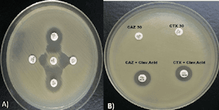
    Due to an increase in community-acquired infections, ESBL-E has become more frequently detected in bacterial cultures.The majority of penicillins, cephalosporins, and aztreonam are rendered inactive by ESBL enzymes. Carbapenems continue to generally be effective against EBSL-E. ESBLs do not render non-lactam agents inactive (e.g., ciprofloxacin, trimethoprim-sulfamethoxazole, gentamicin). However, organisms with ESBL genes frequently have extra genes or gene mutations that cause resistance to a variety of antibiotics. 


     Although ESBL genes can be found in any Gram-negative organism, Escherichia coli, Klebsiella pneumoniae, Klebsiella oxytoca, and Proteus mirabilis have the highest rates of occurrence. The most widespread CTX-M enzymes are CTX-M-15. Other ESBLs with distinctive hydrolyzing properties exist in addition to CTX-M enzymes, such as variants of narrow-spectrum TEM and SHV -lactamases with amino acid substitutions, though they have undergone less thorough clinical testing. Most clinical microbiology laboratories don't conduct routine EBSL testing. Although this threshold has limitations with specificity because organisms that are not susceptible to ceftriaxone for reasons other than ESBL production may be mistakenly assumed to be ESBL-producers, non-susceptibility to ceftriaxone (i.e., ceftriaxone minimum inhibitory concentrations [MICs] 2 mcg/mL) is frequently used as a proxy for ESBL production.

CRE :

      Members of the Enterobacterales order who are resistant to at least one carbapenem antibiotic or who produce a carbapenemase enzyme are known as CRE, according to the CDC .Resistance to at least one carbapenem other than imipenem is necessary for bacteria that are inherently resistant to imipenem (such as Proteus spp., Morganella spp., and Providencia spp.). The pathogens that cause CRE can be roughly divided into those that produce carbapenemase and those that do not into groups with different potential mechanisms of resistance. The amplification of non-carbapenemase -lactamase genes (other than carbapenemase genes) with concurrent outer membrane porin disruption may cause CRE that do not produce carbapenemase. 35% to 59% of CRE cases in the US are caused by isolates that produce carbapenemase.

    K. pneumoniae carbapenemases (KPCs), which can be produced by any Enterobacterales, are the most prevalent carbapenemases. The Verona integron-encoded metallo-lactamases (VIMs), imipenem-hydrolyzing metallo-lactamases (IMPs), and oxacillinases (such as OXA-48-like) are additional notable carbapenemases that have been discovered . Making treatment decisions requires having knowledge of whether a CRE clinical isolate produces carbapenemase and, if so, what kind of carbapenemase is produced.

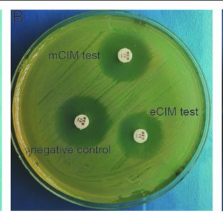
    CRE that produce carbapenemase and those that do not can be distinguished by phenotypic tests like the modified carbapenem inactivation method and the Carba NP test. Molecular analysis can pinpoint particular carbapenemase families (e.g., differentiating a KPC from an OXA-48-like carbapenemase). A small number of clinical microbiology laboratories perform carbapenemase phenotypic and/or genotypic testing, but most of the international bodies strongly encourage all clinical microbiology laboratories to pursue carbapenemase testing in order to guide the best possible treatment choices. The following treatment suggestions for CRE infections presuppose that preferred and alternative antibiotics have shown in vitro activity.

MDR/DTR Pseudomonus aeruginosa:

 Penicillins, cephalosporins, fluoroquinolones, aminoglycosides, and carbapenems are among the antibiotic classes for which P. aeruginosa susceptibility is typically anticipated. MDR P. aeruginosa is defined as P. aeruginosa not susceptible to at least one antibiotic in at least three antibiotic classes . The idea of "difficult-to-treat" resistance was put forth in 2018. DTR is defined  as P. aeruginosa that does not exhibit sensitivity to any of the following drugs: piperacillin-tazobactam, ceftazidime, cefepime, aztreonam, meropenem, imipenem-cilastatin, ciprofloxacin, and levofloxacin.

Multidrug-resistant P. aeruginosa, also known as DTR-P. aeruginosa, typically arises from the interaction of several complex resistance mechanisms, such as reduced expression of outer membrane porins (OprD), increased production of AmpC enzymes, increased activity of efflux pumps, and mutations in targets of the penicillin-binding protein . Carbapenemase production is a rare cause of carbapenem resistance in P. aeruginosa, but it has been found in up to 20% of carbapenem-resistant P. aeruginosa worldwide.


Table . Suggested dosing of antibiotics for the treatment of infections caused by antimicrobial- resistant organisms

Reference : Infectious Disease Society of America (IDSA)


Agent

Adult Dosage

(assuming normal renal and liver function )


Target Organisms 

Amikacin

Cystitis: 15 mg/kg/dose  IV once

All other infections: 20 mg/kg/dose  IV x 1 dose, subsequent doses and dosing interval based on pharmacokinetic evaluation

ESBL-E, AmpC-E, CRE, DTR-

P. aeruginosa

Ampicillin-sulbactam

9 g IV q8h over 4 hours OR 27 g IV q24h as a continuous infusion

For mild infections caused by CRAB isolates susceptible to ampicillin-sulbactam, it is reasonable to administer 3g IV q4h

particularly if intolerance or toxicities preclude the use of higher dosages.

CRAB

Cefepime

Cystitis: 1 g IV q8h

All other infections: 2 g IV q8h, infused over 3 hours

AmpC-E

Cefiderocol

2 g IV q8h, infused over 3 hours

CRE, DTR-P. aeruginosa, CRAB, S. maltophilia

Ceftazidime- avibactam

2.5 g IV q8h, infused over 3 hours

CRE, DTR-P. aeruginosa

Ceftazidime- avibactam and aztreonam

Ceftazidime-avibactam: 2.5 g IV q8h, infused over 3 hours

PLUS

Aztreonam: 2 g IV q8h, infused over 3 hours, administered at the same time as ceftazidime-avibactam, if possible

Metallo-β-lactamase- producing CRE, S. maltophilia

Ceftolozane- tazobactam

Cystitis: 1.5 g IV q8h, infused over 1 hour

All other infections: 3 g IV q8h, infused over 3 hours

DTR-P. aeruginosa

Ciprofloxacin

ESBL-E or AmpC infections: 400 mg IV q8h-q12h OR 500 – 750 mg PO q12h

ESBL-E, AmpC-E

Colistin

Refer to international consensus guidelines on polymyxins 

CRE cystitis, DTR-P. aeruginosa cystitis, CRAB cystitis

Eravacycline

1 mg/kg/dose IV q12h

CRE, CRAB

Ertapenem

1 g IV q24h, infused over 30 minutes

ESBL-E, AmpC-E

Fosfomycin

Cystitis: 3 g PO x 1 dose

ESBL-E. coli cystitis


Agent

Adult Dosage

(assuming normal renal and liver function )


Target Organisms 

Gentamicin

Cystitis: 5 mg/kg/dose  IV once

All other infections: 7 mg/kg/dose  IV x 1 dose, subsequent doses and dosing interval based on pharmacokinetic evaluation

ESBL-E, AmpC-E, CRE,

DTR-P. aeruginosa

Imipenem-cilastatin

Cystitis (standard infusion): 500 mg IV q6h, infused over 30 minutes

All other ESBL-E or AmpC-E infections: 500 mg IV q6h, infused over 30 minutes

All other CRE and CRAB infections: 500 mg IV q6h, infused over 3 hours

ESBL-E, AmpC-E, CRE, CRAB

Imipenem-cilastatin- relebactam

1.25 g IV q6h, infused over 30 minutes

CRE, DTR-P. aeruginosa

Levofloxacin

750 mg IV/PO q24h

ESBL-E, AmpC-E, S.

maltophilia

Meropenem

Cystitis (standard infusion): 1 g IV q8h, infused over 30 minutes

All other ESBL-E or AmpC-E infections: 1-2 g IV q8h, infused over 30 minutes

All other CRE and CRAB infections: 2 g IV q8h, infused over 3 hours

ESBL-E, AmpC-E, CRE, CRAB

Meropenem- vaborbactam

4 g IV q8h, infused over 3 hours

CRE

Minocycline

200 mg IV/PO q12h

CRAB, S. maltophilia

Nitrofurantoin

Cystitis: Macrocrystal/monohydrate (Macrobid®) 100 mg PO q12h

Cystitis: Oral suspension: 50 mg PO q6h

ESBL-E cystitis, AmpC-E cystitis

Plazomicin

Cystitis: 15 mg/kg IV x 1 dose

All other infections: 15 mg/kg IV x 1 dose, subsequent doses and dosing interval based on pharmacokinetic evaluation

ESBL-E, AmpC-E, CRE,

DTR-P. aeruginosa

Polymyxin B

Refer to international consensus guidelines on polymyxins 

DTR-P. aeruginosa, CRAB

Tigecycline

200 mg IV x 1 dose, then 100 mg IV q12h

CRE, CRAB, S. maltophilia


Agent

Adult Dosage

(assuming normal renal and liver function )


Target Organisms 

Tobramycin

Cystitis: 5 mg/kg/dose IV x 1 dose

All other infections: 7 mg/kg/dose IV x 1 dose; subsequent doses and dosing interval based on pharmacokinetic evaluation

ESBL-E, AmpC-E, CRE,

DTR-P. aeruginosa

Trimethoprim- sulfamethoxazole

Cystitis: 160 mg (trimethoprim component) IV/PO q12h

Other infections: 8-12 mg/kg/day (trimethoprim component) IV/PO divided q8-12h (consider maximum dose of 960 mg trimethoprim component per day)

ESBL-E, AmpC-E, S.

maltophilia

AmpC-E: AmpC β-lactamase-producing Enterobacterales; CRAB: Carbapenem-resistant Acinetobacter baumannii; CRE: Carbapenem-resistant Enterobacterales; DTR-P. aeruginosa: Pseudomonas aeruginosa with difficult-to-treat resistance; E. coli: Escherichia coli; ESBL-E: Extended-spectrum β-lactamase-producing Enterobacterales; IV: Intravenous; MIC: Minimum inhibitory concentration; PO: By mouth; q4h: Every 4 hours; q6h: Every 6 hours; q8h: Every 8 hours; q12h: Every 12 hours; q24h: Every 24 hours; S. maltophilia: Stenotrophomonas maltophilia

Tuesday 21 February 2023

Best Antibiotic to Treat UTI in Female

    
     Females in their reproductive years 15-45 often suffer from acute UTI due to sexual activities, short urethra etc. Even UTIs are not uncommon in pregnancy, in which Staphylococcus saprophyticus is often the culprit organism along with other Enterobacteriaceae and Non Fermenters Gram Negative Rods. 

      Patient presents to the doctor with complaints of difficulty in micturition, increased frequency, lower abdominal pain , febrile paroxysmal spikes with chills & rigor , nausea etc. Routine urine microscopy will often reveal pus cells >20/hpf with or without RBCs presence of which may denote upper UTI or pyelonephritis. Urine Culture sample is asked before initiation of 1st dose of emperical antibiotic so that antibiotics doesnot hamper the growth of offending organism thereby limiting its identification and related antibiogram. 

   Now comes the talk of the day ..
Which is best antibiotic to select in UTI in females ? 
     Nitrofurantoin has been the drug of choice in acute uncomplicated cystitis in females as well as children. It's renal tubular secretion is maximum and thereby it attains maximum peak concentration in urine producing optimal therapeutic outcome. Dose is 100 mg twice daily x 7 days for adult females. Common side effects include metallic taste, nausea vomiting, abdominal cramps. 
      Recently Fosfomycin has emerged as a    wonderful drug in treating UTIs in female as it comes as single dose 3 gm sachet to be consumed with 1 glass water which produces execllent therapeutic effect against most of the uropathogens like E.coli, Klebsiella, Proteus etc. Being a Category B drug it is considered safe in pregnancy as well. 
      However, the final antibiotic should always be selected once the culture sensitivity report ( with data of Minimum Inhibitory Concentration/MIC) is available.
Based on antibiogram either an escalating or deescalating approach is adopted to bring out optimal therapeutic outcome following principles of antibiotic stewardship. 

Tuesday 14 February 2023

No New COVID -19 variants emerged in China in 2022 : International Forum

      
     An investigation published in The Lancet found that the recent rise in infections in China following the suspension of the nation's zero-COVID policy did not involve the emergence of any new COVID-19 variations.


    The Lancet reported that a genome analysis of 413 new infections in Beijing since the stringent pandemic safeguards were loosened indicates they were all brought on by pre-existing strands.

      The Lancet reported that between November 14 and December 20, 2022, "more than 90% of local infections in Beijing contained Omicron sub-variants BA.5.2 or BF.7." The majority of imported cases during that time period involved variations from those that were prevalent in Beijing.

      According to reports, China abandoned its zero-COVID policies in December. Targeted lockdowns, massive testing, and quarantines were some of them. The Lancet reported that there have been an increase in cases, prompting fears that new variations may appear.


Tuesday 7 February 2023

Is Fungal infection making your skin itchy ? A comprehensive synopsis

Itchy skin can be attributed to different etiological agents. Amongst those the most common is Dermatophytes which implies superficial fungal skin infection. These Dermatophytes belong to the genera Trichophyton, Epidermophyton and Microsporum. Interestingly, these three genera in order do not affect Skin, Hair & Nail respectively. Different terminologies have been coined to denote site specific involvement like Tinea Corporis( Torso), Tinea Capitis( Scalp), Tinea Cruris( Groin) , Tinea Pedis( Foot) , Tinea Mannum ( Fingers, Hand etc). The most consistent risk factor for Tinea remains poor personal hygiene, moisture due to excessive sweats, tight fitting garments , immune compromised state leading to altered host response like Diabetes Mellitus, Haematological Malignancy, patients receiving chemotherapy, DMARDs, immunomodulatory agents etc. 
Tinea spread through direct contact with infected humans or animals as well as contaminated fomites or vegetative matters. No age group is spared however there is a slight preponderance amongst post pubertal children and young adults probably due to physiological increase in sebum secretion secondary to androgen surge.
      It has been seen that most of the time clinical features of tinea e.g. maculopapular scaly erethamatous lesions with or without central clearing considerably overlap with other dermatological conditions and pose a challenge to clinch the diagnosis. The following chart illustrates the in & out of tinea corporis in an easy to understand format.

So here we go..


Tinea Corporis -


Clinical Features 
Red scaly margin 
Papules & pustules
Mild prutitis
Asymmetrical lesions


Clinical variants 

  ◇Kerion common in children and associated with scalp abscess


◇ Majocchi granuloma common in females around hair & hair follicles of legs


◇ Tinea incognito - Steroid modified tinea mainly found over trunks, limbs etc.


■Clinical Microbiology Lab Diagnosis


10% KOH for morphological identification of fungal etiology with seeding on SDA, PDA for culture confirmation with Fungal AST


Vitek 2 Compact can be employed for Fungal AST with MIC from pure culture growth from SDA/ PDA


Differential Diagnosis of Tinea Corporis:

a) Discoid eczema ( nummular)


Less likely to have central clearing with more confluent scales 

b) Urticaria 
     Erethematous, pruritic with erethamatous wheal 

c) Contact Dermatitis


Well demarcated, erethematous lesion localised to the area of contact

d) Erethema marginatum


Flat erethamatous

e) Atopic Dermatitis 


Highly pruritic, lichenification in chronic lesions and also chronically relapsing

f) Plaque psoriasis


Annular , erethematous round or oval, pruritic plaques. Family History is positive. Nail pitting , arthritis , uveitis

g) Pityriasis rosea


Herald patch progressing to generalised rash 


h) Secondary syphillis 


Diffuse pattern, Symmetrical round or oval, pink -reddish macules 


i) Lichen Planus 


Pruritic papules/plaques

We hope to see you with another interesting Medical blog. Till then eat healthy, sleep well and see you soon ! 


Thursday 2 February 2023

Rotaviral Diarrhoea in Children - A Detail Analysis

The present study entitled, “A study of Molecular Epidemiology of Rotavirus Gastroenteritis among children under Five Years attending NEIGRIHMS, Shillong” was conducted in the Department of Microbiology, NEIGRIHMS from January 2017 to December 2018 with the following objectives: -
1. Understanding the incidence of Rotavirus Gastroenteritis among the children below five years with acute diarrhea attending NEIGRIHMS
2. Molecular characterization of Rotaviruses associated with acute gastroenteritis in children under
five years
3. Study of the clinico-epidemiological profile of Rotaviral Gastroenteritis in children under five years
4. Understanding the correlation between clinical and epidemiological conditions with circulating genotypes of rotaviruses
 In this study, a total of 55 cases of diarrhea fulfilling the case definition were included. Stool samples were collected and each sample was immediately aliquoted up to 3 collection tubes with
proper levelling. 1st set was kept at 2-4 degree Celsius for Rotavirus Group A antigen-based ELISA and others were for kept at -80 degree Celsius for studying the molecular epidemiological pattern
by SDS-PAGE analysis and nested RT-PCR. Childhood diarrhea due to rotavirus can cause significant mortality and morbidity among under five children but unfortunately most of the time
it remains undiagnosed. Moreover, enormous genomic diversity of rotavirus strains is seen across different geographical regions which can challenge the efficacy of currently available rotavirus
vaccines.

The summary of the results and observations made in our study are as follows-
•Amongst a total of 55 under five children, the most common age group that presented to the hospital was 7-9 months which included 13 (23.63%) children. •Rotavirus infection was found to be present in 22(40%) out of 55 total cases. •The highest 9(40.9%) positivity was
again seen in the age group of 7-9 months followed by 4(18.18%) children in the age group of 4-6 months.
•Statistically significant gender specific preponderance was not seen in rotavirus infected children.
•Amongst 22 Rotavirus positive cases, majority 15 (68.18%) was from Meghalaya and Assam 4 (18.18%). Urban and rural distribution of rotavirus positive cases was not found to be significant. Therefore, the present study reaffirms the current concept that Rotavirus is a ‘Democratic Virus’ which can infect children irrespective of the socioeconomic
background of their parents and families.
•In our study, Rotavirus gastroenteritis was found to be most common during the winter months with low temperature and relative humidity. 19(86.36%) rotavirus gastroenteritis cases presented during the months of January to March followed by 3(13.63%) cases who presented between October to December.
•Diarrhea (90%), vomiting (95.45%), fever (90.9%), dehydration’ (90.9%), irritability
(100%), lethargy (100%), were seen as constant clinical features in all the rotavirus gastroenteritis children and found to be statistically significant. 1(4.5%) rotavirus infected child had two episodes of convulsions and loss of consciousness. Dyselectrolytemia in the
form of Hypernatremia (Na+ >145 meq/L) was seen in 16 (72.72%) children while
hypokalemia (K+ <3.5 meq/L) was present in 6(27.27%) children out of 22 Rotavirus
positive gastroenteritis cases. Both these findings were statistically significant.
•This study showed that exclusive breastfeeding per se till the age of 6 months does not confer any overall protection against rotavirus infection.
• A total of 55 stool samples were subjected to sandwich ELISA for Rotavirus Antigen.Rotavirus antigen was positive in 22 (40%) samples. All the ELISA positive samples were subjected to SDS-PAGE electrophoresis in which 11(50%) samples were electropherotyped and characteristic 4:2:3:2 pattern of Rotavirus dsRNA was
demonstrated. All these 11 samples which were electrophoresed showed ‘long
electropherotype’ of Rotaviral dsRNA. ‘Short electropherotype’ of rotavirus was not detected in our study based on the relative mobility pattern of the 11th segment of dsRNA.
• There was no statistically significant age, gender and clinical feature specific difference in the distribution of ‘long electropherotypes’ in our study. No concrete comment could be made in our
study regarding the seasonal distribution of ‘long electropherotype’ of rotavirus.
•All 22 Rotavirus ELISA positive stool samples were subjected to nested RT-PCR for G and P genotyping irrespective of the results obtained in SDS-PAGE electrophoresis.
•Amongst a total of 22 Rotavirus ELISA positive samples, first round of nested RT-PCR for G genotyping gave VP7 amplicon(1062bp) in 11(50%) samples. G genotype was successfully assigned to all these 11 samples (50%) in second round amplification of nested RT-PCR using G- genotype specific primers for G1, G2, G3, G4, G9. All these 11 samples (100%) revealed the presence of G1 genotype. Rest of the 11 samples remained negative in both first and second rounds of nested RT- PCR.

  First round of nested RT-PCR on 22 Rotavirus ELISA positive samples for P genotyping showed VP4 amplicon(663bp) in 10(45.45%) samples. Rest of the 12 samples failed to yield VP4 amplicon (663bp) in our study. Second round of nested RT-PCR was done on all 22 samples using P-genotype specific primers for P[4],P[6],P[8],P[9],P[10]. Amongst 10 samples which gave VP4 amplicons in first round amplification, P [8] genotype was detected in 7 samples (70%) and P [4] was detected in 3 samples (30%). Remaining 12 samples negative in first round, also failed to demonstrate any P-genotype specific amplicon in second round of nested RT-PCR.
•Amongst different combination of G and P genotypes, the most common genotype found in our study was G1P [8] (38.46%) followed by G1P [4] (23.07%).
•Three samples (23.07%) with G1 genotype remained untypeable for P genotypes (VP4 gene).Two samples (15.38%) with P [8] genotype were untypeable for G-Genotype.
  
    Amongst 22 ELISA positive samples, 11 remained untypeable for both VP7 and VP4 genes.
•Definite remarks could not be made regarding the correlation of demographic and clinical features with different G and P genotypes of rotavirus found in our study.
•This study highlighted the different genotypes of rotavirus presently circulating amongst the under five years age children from different north eastern states attending NEIGRIHMS, Shillong.
     
      In our study we observed that amongst 55 under five years age children suffering from diarrhea ,22 (40%) came positive for Rotavirus Group A Antigen by sandwich ELISA. The most common clinical features in these 22 rotavirus infected cases were diarrhea (90.9%), vomiting(95.45%), ‘some dehydration’ (90.09%), fever (90.9%).
       Molecular analysis of these 22 ELISA positive samples was done by SDS-PAGE
electrophoresis in which 11 samples (50%) were electropherotyped and all of them revealed ‘long electropherotype’ pattern of rotavirus dsRNA. Nested RT-PCR carried out on 22 rotavirus ELISA positive samples successfully assigned G -genotype to 11 samples and P- genotype to 10 samples.
All these 11 samples revealed G1 genotype while amongst 10 samples for P-genotyping P[8] was seen in 7 (70%) samples and P[4] in rest 3 (30%) samples. Analysis of different G and P genotype
combinations was also done in our study. The most common genotype combination found was G1P[8] ( 38.46% ) followed by G1 P[4] (23.07%). Rotavirus with genotypes G1P[8] and G1P[4] were found to be consistently associated with diarrhea (100%), vomiting (100%), ‘some
dehydration’(100%), lethargy (100%) and irritability (100%).
     Our study successfully revealed one type of G -Genotype (G1) and two types of P-Genotype (P[4], P[8] ) with the combination of G1P [8], G1P[4] circulating amongst the under five children with rotaviral gastroenteritis attending NEIGRIHMS Shillong .The outcome of our study will be helpful for better understanding of the detail molecular epidemiology of the circulating genotypes of rotaviruses in this region with inclusion of larger sample size for designing the appropriate strategy in control and prevention of Rotavirus gastroenteritis among under five years age children. Further in-depth study in this regard will also help in identifying the appropriate vaccine candidate for development of safe and effective vaccine against rotavirus associated childhood diarrhea.