Showing posts with label Infectious Diseases. Show all posts

Sunday 10 March 2024

 **Diagnosis and Treatment of Sepsis**


By Dr. Koushik Debnath, MD (Clinical Microbiology & Infectious Diseases)


Sepsis is a life-threatening condition that arises when the body's response to infection causes widespread inflammation. As a specialist in Clinical Microbiology & Infectious Diseases, I am deeply committed to raising awareness about the diagnosis and treatment of sepsis, a condition that affects millions worldwide each year.


**Diagnosis:**


Early recognition of sepsis is crucial for successful treatment and improved outcomes. Clinicians rely on a combination of clinical assessment, laboratory tests, and imaging studies to diagnose sepsis accurately.


1. **Clinical Assessment:** Clinicians assess patients for signs and symptoms of infection and systemic inflammation. These may include fever, rapid heart rate, elevated respiratory rate, altered mental status, and low blood pressure.


2. **Laboratory Tests:** Blood cultures are essential for identifying the causative organism and guiding antibiotic therapy. Additionally, biomarkers such as C-reactive protein (CRP) and procalcitonin (PCT) can help in assessing the severity of the infection and monitoring the response to treatment.


3. **Imaging Studies:** Imaging modalities such as chest X-rays, ultrasound, and computed tomography (CT) scans may be performed to identify the source of infection and assess for complications such as pneumonia or intra-abdominal abscesses.


**Treatment:**


The management of sepsis requires a multi-disciplinary approach involving early recognition, aggressive resuscitation, and targeted antimicrobial therapy.


1. **Fluid Resuscitation:** Prompt administration of intravenous fluids is essential to restore perfusion and optimize organ function. Clinicians typically initiate fluid resuscitation with crystalloids such as isotonic saline or lactated Ringer's solution.


2. **Antimicrobial Therapy:** Empirical antimicrobial therapy should be initiated as soon as sepsis is suspected, based on the likely source of infection and local antibiotic resistance patterns. Once blood cultures are obtained, therapy can be adjusted based on culture results and antimicrobial susceptibility testing.


3. **Vasopressor Support:** Patients with septic shock may require vasopressor medications such as norepinephrine or vasopressin to maintain adequate blood pressure and perfusion to vital organs.


4. **Supportive Care:** Supportive measures such as mechanical ventilation, renal replacement therapy, and nutritional support may be necessary for patients with severe sepsis or septic shock.


5. **Source Control:** Definitive management of sepsis often involves identifying and eliminating the source of infection through surgical or interventional procedures, such as drainage of abscesses or removal of infected tissue.


**Conclusion:**


Sepsis remains a significant cause of morbidity and mortality worldwide, highlighting the importance of early recognition and aggressive management. As clinicians, it is our responsibility to remain vigilant for signs of sepsis, initiate timely interventions, and collaborate closely with other healthcare providers to optimize patient outcomes. Through ongoing education and research, we can continue to improve our understanding and management of this complex condition, ultimately saving lives and reducing the burden of sepsis on individuals and healthcare systems alike.

Thursday 1 June 2023

Unveiling HMPV: A Comprehensive Guide to the Spreading Virus in the US


Introduction:

HMPV, short for Human Metapneumovirus, is a virus that is currently spreading in the United States. It's important to understand what this virus is and how it can affect us. In this article, we will provide you with an easy-to-understand explanation of HMPV and its key characteristics.


1. What is HMPV?

HMPV is a type of virus that primarily affects the respiratory system. It belongs to the same family as the common cold and respiratory syncytial virus (RSV). It was first identified in 2001 and has since been recognized as a common cause of respiratory infections, especially in young children and older adults.


2. How does HMPV spread?

HMPV spreads from person to person through respiratory droplets when an infected individual coughs or sneezes. These droplets can then be inhaled by nearby individuals, leading to infection. It can also spread by touching surfaces contaminated with the virus and then touching the face.


3. What are the symptoms?

The symptoms of HMPV are similar to those of other respiratory infections. They may include cough, runny nose, sore throat, fever, and difficulty breathing. In severe cases, it can lead to pneumonia or bronchiolitis, particularly in young children or individuals with weakened immune systems.


4. Who is at risk?

While HMPV can affect people of all ages, it is more likely to cause severe illness in infants, young children, older adults, and individuals with underlying health conditions. These individuals should take extra precautions to avoid exposure to the virus.


5. How can HMPV be prevented?

Preventing the spread of HMPV involves practicing good hygiene and taking precautions to minimize exposure. Some preventive measures include:

- Washing hands frequently with soap and water for at least 20 seconds.

- Using hand sanitizers if soap and water are not readily available.

- Covering the mouth and nose with a tissue or elbow when coughing or sneezing.

- Avoiding close contact with individuals showing symptoms of respiratory infection.

- Disinfecting frequently-touched surfaces regularly.


6. Is there a specific treatment ?

Rest with supportive care, such as getting plenty of rest, staying hydrated, and using over-the-counter medications to alleviate symptoms. In severe cases, hospitalization may be required.


7. When should I seek medical attention?

If you or a loved one experience severe symptoms, such as persistent high fever, difficulty breathing, or worsening cough, it is advisable to seek medical attention promptly. Healthcare professionals can provide a proper diagnosis and offer appropriate treatment recommendations.


Conclusion:

HMPV is a respiratory virus that is spreading in the United States. It can cause symptoms similar to the common cold or more severe respiratory infections. By practicing good hygiene, taking preventive measures, and seeking medical attention when necessary, we can minimize the spread and impact of HMPV. Stay informed and take care of your health and well-being.

Wednesday 31 May 2023

Real-Time PCR: Unveiling the Importance of CT Value in Infectious Disease Treatment


Introduction:


      Infectious diseases continue to pose a significant threat to public health globally. The ability to rapidly and accurately diagnose these diseases is crucial for effective treatment and containment. Real-time polymerase chain reaction (PCR) has revolutionized the field of infectious disease diagnostics, allowing for precise and timely detection of pathogens. One of the key parameters derived from real-time PCR analysis is the CT value, which holds immense importance in guiding treatment decisions and monitoring disease progression. This article aims to explore the concept of real-time PCR and delve into the significance of the CT value in the context of infectious disease treatment.


Understanding Real-Time PCR:


       Polymerase chain reaction (PCR) is a laboratory technique used to amplify a specific segment of DNA or RNA from a complex mixture. Traditional PCR involves multiple cycles of amplification and requires a separate step for detecting the amplified product post-amplification. Real-time PCR, also known as quantitative PCR (qPCR), is an advanced version that allows the detection of amplified DNA or RNA in real-time as the reaction progresses. This real-time monitoring is achieved by incorporating fluorescent dyes or probes into the reaction mixture.


     Real-time PCR is a highly sensitive and specific technique that enables the rapid identification and quantification of nucleic acids. It is widely employed for diagnosing various infectious diseases, including viral, bacterial, and fungal infections. By targeting specific regions of the pathogen's genetic material, real-time PCR can confirm the presence of the infectious agent with high accuracy.


The Significance of the CT Value:


      The CT value, also referred to as the cycle threshold or quantification cycle (Cq), is a crucial parameter derived from real-time PCR analysis. It represents the cycle number at which the fluorescence signal generated by the amplification reaches a specific threshold level. The CT value is inversely proportional to the amount of target nucleic acid initially present in the sample. A lower CT value indicates a higher initial target concentration, while a higher CT value suggests a lower initial target concentration.


      In the context of infectious disease treatment, the CT value holds significant importance for several reasons:


1. Diagnostic Potential: 

      The CT value provides a quantitative measure of the pathogen's load in a clinical sample. By comparing the CT value obtained from a patient's sample to established reference ranges, clinicians can determine whether an infection is present, aiding in accurate diagnosis.


2. Monitoring Disease Progression:

      During the course of an infection, the CT value can serve as an indicator of disease progression or response to treatment. A decreasing CT value over time suggests a reduction in pathogen load, indicating successful treatment. Conversely, an increasing CT value may indicate treatment failure or the emergence of drug resistance.


3. Treatment Guidance: 

      The CT value can help guide treatment decisions, particularly in the case of viral infections. Different viral infections have varying levels of virulence and response to antiviral medications. By monitoring the CT value over time, clinicians can assess the effectiveness of antiviral therapy and make adjustments accordingly.


4. Contagiousness Assessment

         The CT value can provide insights into the contagiousness of an individual infected with a particular pathogen. Lower CT values indicate higher viral loads and suggest that the patient may be more contagious. This information can assist in implementing appropriate infection control measures to limit the spread of the disease.


5. Prognostic Indicator:

       In some cases, the CT value has been correlated with the severity of the disease and patient outcomes. Studies have shown that a higher CT value at the time of diagnosis may be associated with a milder course of illness, while a lower CT value may indicate a higher risk of complications or adverse outcomes.


Conclusion:


      Real-time PCR has revolutionized infectious disease diagnostics, providing rapid and accurate detection of pathogens.

Saturday 15 April 2023

Hepatitis B - A Vaccine preventable serious Liver Infection

Acute hepatitis B virus infection:


Thirty percent of patients with acute hepatitis B develop icteric hepatitis, while roughly seventy percent have subclinical or anicteric hepatitis. Patients with underlying liver disease or other hepatitis viruses may have a more severe case of the disease.

       One to six months pass during the incubation phase. During the prodromal stage, a syndrome resembling serum sickness may appear, followed by constitutional symptoms, anorexia, nausea, jaundice, and discomfort in the right upper quadrant. In most cases, the symptoms and jaundice go away after one to three months. A rare condition, cute liver failure affects 0.1 to 0.5 percent of patients.

Acute hepatitis with HBsAg positivity can also be diagnosed as: 

(1) Acute hepatitis B

(2) Chronic hepatitis B exacerbations, such as acute hepatitis brought on by drugs and other toxins in a chronic hepatitis B infected person, reactivation of chronic hepatitis B, or superinfection of a chronic hepatitis B infection with hepatitis C, A, E, or D virus.

       Alanine and aspartate aminotransferase levels (ALT and AST) are elevated during the acute phase of acute hepatitis B; values up to 1,000 to 2,000 IU/L are typically seen during this phase, with ALT being higher than AST. The serum levels of lactic dehydrogenase and alkaline phosphatase are typically only slightly elevated (less than threefold). Both the direct and indirect fractions of the bilirubin are variablely elevated. Patients with an-icteric hepatitis may have normal serum bilirubin levels. Except in cases of chronic, severe disease, serum albumin rarely decreases. The best prognosis predictor is the prothrombin time.

        Normalisation of serum aminotransferases takes one to four months to happen in patients who make a full recovery. Persistent elevation of serum ALT for more than six months may indicate progression to chronic hepatitis.

Chronic hepatitis B virus infection:

     The age of infection has a major impact on how quickly acute hepatitis B turns into chronic hepatitis B. The rate is roughly 90% for infections acquired during pregnancy, 20% to 50% for infections acquired between ages one and five, and less than 5% for infections acquired as adults. Hepatitis B virus infection is referred to as chronic if HBsAg is persistently positive for more than six months.

    A thorough history and physical examination with a focus on determining the severity of underlying liver disease and determining treatment eligibility should be performed on people with chronic hepatitis B. Alcohol use, family history of HBV infection, liver disease, and liver cancer, history of complications that would suggest underlying cirrhosis (such as ascites, hematemesis, and mental status changes), and other factors, such as underlying cardiopulmonary disease, past or present psychiatric issues, autoimmune diseases, and other co-morbid conditions, should all be highlighted in the history. Evaluation of advanced liver disease stigmata, such as spider angiomata, palmar erythema, splenomegaly, jaundice, or caput medusa, should be part of a physical examination. Clinicians should be aware, though, that the absence of any one of these findings does not rule out the possibility of underlying cirrhosis. 
    Patients diagnosed with Chronic Hepatitis B should also be mandatorily  screened for HIV and Hepatitis C due to common modes of transmission.

Laboratory Diagnosis : 
   Blood is the preferred specimen, and 3-5 ml of venous blood needs to be drawn and placed in a sterile, dry, and labelled vial. Avoid hemolyzed samples because they might make it difficult for tests to detect markers accurately.
• Within 4 hours of collection, serum from clotted blood should be removed and stored at -20°C to -70°C to prevent the degradation of viral nucleic acid in the specimen.
• Serum samples can be stored for a maximum of 7 days at 4-8°C and  long term storage beyond 7 days should be done at -80 °C. 

Serological markers for Chronic Hepatitis B infection:

1) Hepatitis B surface antigen (HBsAg): 

     After infection, HBV DNA is the first serological marker, followed by HBsAg. Even before the elevation of liver enzymes and the start of a clinical illness, the antigen is detectable. Typically, it goes away 2 months after the start of the clinical illness, but in some cases, such as with chronic infections, it can persist for up to 6 months. Therefore, if this test results in a positive result, the patient is likely contagious, and if it results in a negative result, chronic infection is usually ruled out.

2) Anti-HBsAg (Antibody to HBV surface antigen):

     This antibody manifests when HBsAg is no longer detectable (antibody to HBV surface antigen).It is a protective antibody that shows immunity to HBV from either a prior infection or vaccination.Anti-HBs antibodies have a protective level of 10 mIU/ml.


3) Anti-HBc ( Antibody to HBV core antigen):

       The earliest antibody marker after infection is the anti-HBcIgM (antibody to HBV core antigen). The anti-HBcIgM is the first antibody marker to be detected in blood, appearing in serum a week or two after HBsAg does. Since the anti-HBcIgG antibody may last a lifetime, it serves as a useful marker for previous HBV infection. Total antibody to HBc indicates past or ongoing infection because IgM anti-HBc is only present in acute infections and is replaced by IgG after six months. IgG anti-HBc is a trustworthy indicator of prior HBV infection because it endures even after anti-HBs titers drop to undetectable levels many years following recovery from HBV infection.

4) HBeAg

     In acute, resolving cases, it typically vanishes within a few weeks after appearing in the blood concurrently with or shortly after HBsAg.The presence of this antigen is also used as a criterion for selecting patients for treatment because it is an indicator of active intrahepatic viral replication and, as a result, indicates that the person is highly contagious.Anti-HBe then appears after its disappearance. In the majority of acute hepatitis B cases, testing for HBeAg is not required for routine diagnostic purposes. Instead, individuals in whom HBeAg is a significant marker of viral replication and correlates qualitatively with more quantitative markers of active replication, such as serum HBV DNA detected using molecular techniques, should have their HBeAg levels tested.

  5)Anti-HBeAg :
     
      It has prognostic implications as the appearance of anti-HBe in acute hepatitis B implies a high likelihood that HBV infection will resolve spontaneously. Anti-HBe's presence in blood denotes low infectivity.

 Serologic markers-caveats:
      
  •  Precore mutants have mutations in the precore region, which abolishes HBeAg production, or in the core promoter region, which downregulates HBeAg production. Despite possibly producing anti-HBe and anti-HBc antibodies, these patients do not produce HBeAg. 
  • The second group of so-called escape mutants (due to mutations in a determinant of S gene preventing them from being neutralised by the anti-HBs) is seen in some infants born to HBeAg positive mothers and in liver transplant patients who have received combined immunisation with anti-HBV immunoglobulin and vaccine. This has no effect on viral replication; in fact, such cases are more difficult to treat and have a higher risk of turning into cirrhosis. 
  • HBeAg and HBsAg may both be suppressed by co-infection with HCV.
  • HBcAb is occasionally the only serological marker that can be found. This could be either  because of Co-infection with HCV or HIV, Remote infection, "Window" period between HBsAg and HBsAb, False positive test result - HBcAb is marker most prone to false positives or  Resolved HBV with diminishing anti-HBs levels.

 Hepatitis B Occult infection (OBI)

      Viral DNA in blood that is in motion without HBsAg being visible. HBV DNA and anti-HBc would be the only remaining markers after anti-HBe disappears.

Molecular Diagnostics :

 HBV DNA (Quantitative): 

    Hepatitis B DNA PCR is a molecular test that can detect the presence of HBV DNA, which is a marker of viral replication and infectivity like HBeAg.Therefore, HBV DNA is used in clinical practise to track therapy and evaluate patient response to it, such as every three months for years when the patient is taking oral agents and every month for six to twelve months if the patient is taking PEG/IFN. The identification of occult HBV infection is another use for it.

Genotyping and Resistance Testing :

      The genotyping is recommended for both epidemiological purposes in the case of an outbreak investigation and for the detection of mutations that confer resistance to antiviral agents. Eight different HBV genotypes (A to H) have been identified through genotyping of patient isolates. Sequencing and hybridization techniques (Line Probe Assay) are used for genotyping.


Treatment of Chronic Hepatitis B: 

Candidates for treatment -

  • Regardless of ALT levels, HBeAg status, or HBV DNA levels, treatment for all adults, adolescents, and kids with CHB and signs of compensated or decompensated cirrhosis should be prioritised.
  • Regardless of HBeAg status, treatment is advised for adults with CHB who do not have cirrhosis but are older than 30 (in particular), have persistently abnormal ALT levels, and show evidence of high-level HBV replication (HBV DNA >20 000 IU/mL).
Whom NOT to Treat :

  • In people without signs of cirrhosis, with persistently normal ALT levels, and with low levels of HBV replication (HBV DNA 2000 IU/mL), regardless of HBeAg status or age, antiviral therapy is not advised and can be postponed.
  • In the absence of HBV DNA testing, treatment can be postponed in HBeAg-positive people 30 years of age or younger with persistently normal ALT levels.
  • All CHB patients need to be monitored regularly, but it's especially important for those who don't currently fit the guidelines for whether or not to receive treatment, to find out if antiviral therapy will ever be necessary to stop the progression of liver disease. These people include those under 30 years old without cirrhosis who have HBV DNA levels greater than 20,000 IU/mL but persistently normal ALT.

Treatment modalities of Chronic Hepatitis B Infection :

  • The NAs with a high barrier to drug resistance (tenofovir or entecavir) are advised for all adults, adolescents, and children aged 12 years or older for whom antiviral therapy is indicated.
  • Tenofovir may be preferred as the medication of choice in women of childbearing age in the event of pregnancy. Pregnancy is not advised when using entecavir.
  • Patients with a risk of developing Entecavir resistance who have taken lamivudine are advised to use tenofovir instead.
  • Children between the ages of 2 and 11 should not take entecavir.
  • Entecavir may be preferred over Tenofovir in Age > 60, history of fragility fractures or osteoporosis; use of other drugs that worsen bone density, altered renal function (eGFR 60 mL/min/1.73 m2, albuminuria > 30 mg/24 hr, moderate dipstick proteinuria, Low phosphate (2.5 mg/dL) or in patients receiving hemodialysis.
  • Drugs with a low barrier to resistance (lamivudine, adefovir, or telbivudine) are available but not advised as they promote drug resistance. Tenofovir alafenamide fumarate (TAF) is the drug of choice in patients with reduced renal function or bone disease bone toxicities, where entecavir is contraindicated.   
      Prior to, during, and after treatment, CHB patients are monitored for disease progression and treatment response.It is advised that the following be checked at least once a year:
  • HBV DNA levels (where HBV DNA testing is available), HBsAg, HBeAg, and ALT levels (and AST for APRI)
  • Non-invasive tests (APRI score, FIB-4, or FibroScan) to determine whether fibrosis has gotten worse or whether cirrhosis has developed in people who didn't have it at the start.
  • If receiving treatment, adherence needs to be checked frequently and at each appointment.
    Given the risk of reactivation, which can also result in severe acute-on-chronic liver injury, all individuals with cirrhosis based on clinical evidence (or adults with an APRI score >2) require lifelong treatment with NAs.
   Hepatitis B treatment should not typically be stopped without consulting medical professionals with the necessary knowledge, who should be found in specialised facilities.
    When deciding to stop therapy, it is important to carefully weigh the financial impact of continuing to pay for medication and monitoring versus the risk of virological relapse, decompensation, and death. Antiviral treatment should not be stopped for any cirrhotic patients due to the possibility of reactivation, which could result in decompensation and death.


Monday 27 March 2023

Scrub Typhus - A potentially life threatening fever ?

 What is scrub typhus ?

    Orientia tsutsugamushi, an obligate intracellular gram-negative bacterium, is the source of the acute febrile illness known as scrub typhus. Although scrub typhus caused by other Orientia species has been reported in Africa, France, the Middle East, and South America, it is believed to be endemic to the tsutsugamushi triangle, which includes Asia, northern Australia, and islands in the Indian and Pacific Oceans. According to a recent systematic review from hospital-based studies in India, scrub typhus was the root cause of 25% of acute undifferentiated febrile illnesses. 


     O. tsutsugamushi transmitted by the bites of infected chiggars( trombiculid mite) has been linked in recent studies to acute encephalitis syndrome (AES) outbreaks in India, particularly in the northern states of Uttar Pradesh, Bihar, West Bengal, and Assam. Outbreaks of AES pose a major public health problem in India, predominantly affecting children. 

Spectrum of Clinical Features-

      All patients who exhibit an acute onset of fever and altered mental status are included in the broad definition of Acute Encephalitis Syndrome used for syndromic surveillance. Typical clinical features include fever with chills & rigor ,headache,bodyaches,muscle cramps,altered sensorium ,lymphadenopathy and rash .


 The pathognomonic classical scrub typhus rash has been described as a dark scab like region at the site of the chiggar bite, popularly known as 'Eschar'.

      The clinical manifestation may be brought on by encephalitis, meningitis, or encephalopathy without CNS invasion, such as in cases of severe systemic infection, metabolic disturbance, or other neurologic complications following the infection. 

Diagnostic Challenges-

        Early diagnosis is essential for starting prompt, targeted treatment, which can lower scrub typhus complications and fatality rates. Due to the symptoms' similarity to those of other tropical infections that are endemic to the region and can also cause AES, such as dengue, chikungunya, malaria, and leptospirosis, clinical diagnosis can be difficult. 

       There are drawbacks to the current microbiological diagnostics for scrub typhus, which are typically based on IgM detection in serum samples or nucleic acid by PCR. IgM can persist for a long time after the onset of acute illness, and it can react with IgM from other cocirculating pathogens. IgM first appears in serum 5–6 days after the onset of illness .Therefore, it is challenging to determine O. tsutsugamushi as the cause in AES patients who also have microbiological evidence of another potential pathogen.


        Patients with suspected neurologic scrub typhus do not undergo routine IgM detection in cerebrospinal fluid (CSF).The immunofluorescence assay has long been regarded as the gold standard in serologic testing, but its use is constrained by its high cost and difficult interpretation. Serologic tests' limitations in terms of cross-reacting and persistent antibodies may be addressed by PCR, but a positive result is only likely to occur during the bacteremia stage of infection .

        Additionally, blood or eschar material are suggested samples for O. tsutsugamushi PCR, whereas it is unknown how sensitive PCR is to CSF.

          More than 50% of patients had anaemia, leukocytosis, thrombocytopenia, transaminitis, hypoalbuminemia, and uremia. Most patients' CSF tests showed lymphocytic pleocytosis and increased protein concentration.

Treatment-

       Doxycycline 100mg twice daily x 10 days has been the drug of choice for most of the patients without having obvious comorbid conditions. Supportive therapy in terms of Paracetamol, Intravenous Fluids etc are often required. Comorbid conditions like Diabetes Mellitus, Pre-existing Nephropathy can lead to AKI like manifestations in selected group of patients. Hence a constant vigilant monitoring of serum creatinine, electrolytes along with CRP/Procalcitonin  may be of importance to optimise the therapeutic outcome. 

Sunday 5 March 2023

Typhoid vaccine price in India - Know why it is worth the cost

 What is Typhoid Fever ?

    A potentially fatal multisystemic infection known as enteric fever, also known as typhoid fever is caused primarily by Salmonella enterica serotype typhi and, to a lesser extent, paratyphi A, B, and C. ICD 10 code for enteric fever is now A01.00. Salmonella are motile enterobacteriaceae that can cause a number of different gastrointestinal infections. Typhoid, which is primarily caused by Salmonella enterica serotype typhi and, to a lesser extent, S enterica serotypes paratyphi A, B, and C, is the most dangerous of these. From a severe septic illness to minor cases of diarrhoea with low-grade fever, it can present in a wide range of ways. Fever, malaise, diffuse abdominal pain, and constipation are characteristics of the classic presentation. Typhoid fever that is left untreated can lead to delirium, obtundation, intestinal bleeding, bowel perforation, and death in less than a month.

    It has grown more and more resistant to antibiotics over time. Extensively drug-resistant typhoid (XDR) was reported in Pakistan in 2016. Azithromycin, carbapenems, and tigecycline are the only antimicrobial classes that are still effective against these strains.

     Conditions of poor sanitation, overcrowding, and social unrest favour the growth of typhoid.  Typhoid fever is still endemic in developing nations, despite the fact that antibiotics have significantly decreased its prevalence in the developed world. It's possible that S paratyphi infections are becoming more common than S typhi infections. This might be brought on by the immunologic immaturity of a particular population as well as the insufficient protection against these pathogens offered by the current Typhoid vaccines. Most commonly, non-Typhoidal strains cause a mild, self-limiting gastroenteritis. Nevertheless Typhoid vaccines play a crucial role both in primary prevention and decreasing the disease severity in affected individuals.

Vaccines for prevention :

Courtesy : Dr Koushik's Medicare 

        Typhoid fever can be prevented by two vaccinations. The first is a live, attenuated (weakened) vaccine, and the second is an inactivated (dead) vaccine. Which typhoid vaccine is ideal for you can be decided with the aid of your doctor. Typhoid vaccination that has been inactivated is given by injection. Children of 2 years of age and older may receive it. At least two weeks prior to departure, one dose is advised. For those who continue to be at danger, repeated doses every two years are advised.

        Oral live typhoid vaccine administration (by mouth). Six years of age and older individuals may receive it. Every other day, one capsule is taken, making a total of four capsules. At least a week before departure, the last dose should be taken.About an hour before meals, each capsule should be taken whole (without chewing) with cold or lukewarm water. For those who continue to be at risk, a booster shot is required every five years. It's crucial to keep live typhoid vaccine capsules chilled (not frozen).

      In developed countries  most of the time routine typhoid immunisation is not advised, although the following situations may warrant administration of typhoid vaccines.

a) Tourists going to regions where typhoid is widespread. (NOTE: Typhoid vaccination is not 100% effective and is not a replacement for exercising caution while choosing foods and beverages.)

b) Individuals in close proximity to a typhoid carrier.

c) Employees in laboratories that handle Salmonella typhi bacterial culture and strains

The typhoid vaccine may be administered concurrently with other shots.

     In 2023 typhoid vaccine price in India is around INR 1500-2500 based on the brands available. You can check the same on : https://www.apollopharmacy.in/medicine/typbar-tcv-pfs-injection-0-5ml

     Typbar TCV Vaccine 0.5 ml. It includes sodium chloride and the purified Vi polysaccharide typhoid vaccine. Typhoid disease is prevented with it. A non-toxic and safe form of the causative bacteria's capsular specimen is injected into the body by Typbar TCV Vaccine, which prompts the body to produce antibodies and confers immunity. The body keeps track of this immune response to ensure that it is ready in the event that these bacteria invade again in the future.

Friday 24 February 2023

Difficult to Treat Infections ? Take a look

     Antibiotics have been a wonderful invention in the evolution of modern medical treatments. The mortality and the morbidity due to different infectious diseases have remarkably gone down over the last many decades. However pathogens have emerged as even more smarter than mankind developing different complex resistance mechanisms to survive the assault of antibiotics thereby rendering treatment of infectious diseases a difficult challenge. Patients with Diabetic Ketoacidosis, Chronic Renal failure, Implants & Prosthesis often land up with sepsis with multi drug resistant organisms ,treatment of which pose a serious challenge to the treating clinicians. To combat this crisis, antibiotic stewardship practice has become the need of the hour which emphasize the rational & judicious use of all available antibiotics. The regulatory authority should be more vigilant in preventing the over the counter dispensing of antibiotics.


     Having said that, all the medical fraternities and academic forums should take initiatives to implement antibiotic stewardship practices in day to day clinical practices. Let us have a quick brush up of the antibiotic resistance mechanisms.   


ESBLs :

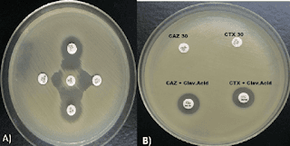
    Due to an increase in community-acquired infections, ESBL-E has become more frequently detected in bacterial cultures.The majority of penicillins, cephalosporins, and aztreonam are rendered inactive by ESBL enzymes. Carbapenems continue to generally be effective against EBSL-E. ESBLs do not render non-lactam agents inactive (e.g., ciprofloxacin, trimethoprim-sulfamethoxazole, gentamicin). However, organisms with ESBL genes frequently have extra genes or gene mutations that cause resistance to a variety of antibiotics. 


     Although ESBL genes can be found in any Gram-negative organism, Escherichia coli, Klebsiella pneumoniae, Klebsiella oxytoca, and Proteus mirabilis have the highest rates of occurrence. The most widespread CTX-M enzymes are CTX-M-15. Other ESBLs with distinctive hydrolyzing properties exist in addition to CTX-M enzymes, such as variants of narrow-spectrum TEM and SHV -lactamases with amino acid substitutions, though they have undergone less thorough clinical testing. Most clinical microbiology laboratories don't conduct routine EBSL testing. Although this threshold has limitations with specificity because organisms that are not susceptible to ceftriaxone for reasons other than ESBL production may be mistakenly assumed to be ESBL-producers, non-susceptibility to ceftriaxone (i.e., ceftriaxone minimum inhibitory concentrations [MICs] 2 mcg/mL) is frequently used as a proxy for ESBL production.

CRE :

      Members of the Enterobacterales order who are resistant to at least one carbapenem antibiotic or who produce a carbapenemase enzyme are known as CRE, according to the CDC .Resistance to at least one carbapenem other than imipenem is necessary for bacteria that are inherently resistant to imipenem (such as Proteus spp., Morganella spp., and Providencia spp.). The pathogens that cause CRE can be roughly divided into those that produce carbapenemase and those that do not into groups with different potential mechanisms of resistance. The amplification of non-carbapenemase -lactamase genes (other than carbapenemase genes) with concurrent outer membrane porin disruption may cause CRE that do not produce carbapenemase. 35% to 59% of CRE cases in the US are caused by isolates that produce carbapenemase.

    K. pneumoniae carbapenemases (KPCs), which can be produced by any Enterobacterales, are the most prevalent carbapenemases. The Verona integron-encoded metallo-lactamases (VIMs), imipenem-hydrolyzing metallo-lactamases (IMPs), and oxacillinases (such as OXA-48-like) are additional notable carbapenemases that have been discovered . Making treatment decisions requires having knowledge of whether a CRE clinical isolate produces carbapenemase and, if so, what kind of carbapenemase is produced.

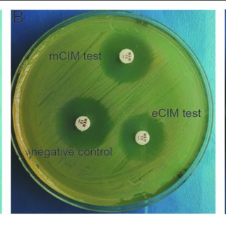
    CRE that produce carbapenemase and those that do not can be distinguished by phenotypic tests like the modified carbapenem inactivation method and the Carba NP test. Molecular analysis can pinpoint particular carbapenemase families (e.g., differentiating a KPC from an OXA-48-like carbapenemase). A small number of clinical microbiology laboratories perform carbapenemase phenotypic and/or genotypic testing, but most of the international bodies strongly encourage all clinical microbiology laboratories to pursue carbapenemase testing in order to guide the best possible treatment choices. The following treatment suggestions for CRE infections presuppose that preferred and alternative antibiotics have shown in vitro activity.

MDR/DTR Pseudomonus aeruginosa:

 Penicillins, cephalosporins, fluoroquinolones, aminoglycosides, and carbapenems are among the antibiotic classes for which P. aeruginosa susceptibility is typically anticipated. MDR P. aeruginosa is defined as P. aeruginosa not susceptible to at least one antibiotic in at least three antibiotic classes . The idea of "difficult-to-treat" resistance was put forth in 2018. DTR is defined  as P. aeruginosa that does not exhibit sensitivity to any of the following drugs: piperacillin-tazobactam, ceftazidime, cefepime, aztreonam, meropenem, imipenem-cilastatin, ciprofloxacin, and levofloxacin.

Multidrug-resistant P. aeruginosa, also known as DTR-P. aeruginosa, typically arises from the interaction of several complex resistance mechanisms, such as reduced expression of outer membrane porins (OprD), increased production of AmpC enzymes, increased activity of efflux pumps, and mutations in targets of the penicillin-binding protein . Carbapenemase production is a rare cause of carbapenem resistance in P. aeruginosa, but it has been found in up to 20% of carbapenem-resistant P. aeruginosa worldwide.


Table . Suggested dosing of antibiotics for the treatment of infections caused by antimicrobial- resistant organisms

Reference : Infectious Disease Society of America (IDSA)


Agent

Adult Dosage

(assuming normal renal and liver function )


Target Organisms 

Amikacin

Cystitis: 15 mg/kg/dose  IV once

All other infections: 20 mg/kg/dose  IV x 1 dose, subsequent doses and dosing interval based on pharmacokinetic evaluation

ESBL-E, AmpC-E, CRE, DTR-

P. aeruginosa

Ampicillin-sulbactam

9 g IV q8h over 4 hours OR 27 g IV q24h as a continuous infusion

For mild infections caused by CRAB isolates susceptible to ampicillin-sulbactam, it is reasonable to administer 3g IV q4h

particularly if intolerance or toxicities preclude the use of higher dosages.

CRAB

Cefepime

Cystitis: 1 g IV q8h

All other infections: 2 g IV q8h, infused over 3 hours

AmpC-E

Cefiderocol

2 g IV q8h, infused over 3 hours

CRE, DTR-P. aeruginosa, CRAB, S. maltophilia

Ceftazidime- avibactam

2.5 g IV q8h, infused over 3 hours

CRE, DTR-P. aeruginosa

Ceftazidime- avibactam and aztreonam

Ceftazidime-avibactam: 2.5 g IV q8h, infused over 3 hours

PLUS

Aztreonam: 2 g IV q8h, infused over 3 hours, administered at the same time as ceftazidime-avibactam, if possible

Metallo-β-lactamase- producing CRE, S. maltophilia

Ceftolozane- tazobactam

Cystitis: 1.5 g IV q8h, infused over 1 hour

All other infections: 3 g IV q8h, infused over 3 hours

DTR-P. aeruginosa

Ciprofloxacin

ESBL-E or AmpC infections: 400 mg IV q8h-q12h OR 500 – 750 mg PO q12h

ESBL-E, AmpC-E

Colistin

Refer to international consensus guidelines on polymyxins 

CRE cystitis, DTR-P. aeruginosa cystitis, CRAB cystitis

Eravacycline

1 mg/kg/dose IV q12h

CRE, CRAB

Ertapenem

1 g IV q24h, infused over 30 minutes

ESBL-E, AmpC-E

Fosfomycin

Cystitis: 3 g PO x 1 dose

ESBL-E. coli cystitis


Agent

Adult Dosage

(assuming normal renal and liver function )


Target Organisms 

Gentamicin

Cystitis: 5 mg/kg/dose  IV once

All other infections: 7 mg/kg/dose  IV x 1 dose, subsequent doses and dosing interval based on pharmacokinetic evaluation

ESBL-E, AmpC-E, CRE,

DTR-P. aeruginosa

Imipenem-cilastatin

Cystitis (standard infusion): 500 mg IV q6h, infused over 30 minutes

All other ESBL-E or AmpC-E infections: 500 mg IV q6h, infused over 30 minutes

All other CRE and CRAB infections: 500 mg IV q6h, infused over 3 hours

ESBL-E, AmpC-E, CRE, CRAB

Imipenem-cilastatin- relebactam

1.25 g IV q6h, infused over 30 minutes

CRE, DTR-P. aeruginosa

Levofloxacin

750 mg IV/PO q24h

ESBL-E, AmpC-E, S.

maltophilia

Meropenem

Cystitis (standard infusion): 1 g IV q8h, infused over 30 minutes

All other ESBL-E or AmpC-E infections: 1-2 g IV q8h, infused over 30 minutes

All other CRE and CRAB infections: 2 g IV q8h, infused over 3 hours

ESBL-E, AmpC-E, CRE, CRAB

Meropenem- vaborbactam

4 g IV q8h, infused over 3 hours

CRE

Minocycline

200 mg IV/PO q12h

CRAB, S. maltophilia

Nitrofurantoin

Cystitis: Macrocrystal/monohydrate (Macrobid®) 100 mg PO q12h

Cystitis: Oral suspension: 50 mg PO q6h

ESBL-E cystitis, AmpC-E cystitis

Plazomicin

Cystitis: 15 mg/kg IV x 1 dose

All other infections: 15 mg/kg IV x 1 dose, subsequent doses and dosing interval based on pharmacokinetic evaluation

ESBL-E, AmpC-E, CRE,

DTR-P. aeruginosa

Polymyxin B

Refer to international consensus guidelines on polymyxins 

DTR-P. aeruginosa, CRAB

Tigecycline

200 mg IV x 1 dose, then 100 mg IV q12h

CRE, CRAB, S. maltophilia


Agent

Adult Dosage

(assuming normal renal and liver function )


Target Organisms 

Tobramycin

Cystitis: 5 mg/kg/dose IV x 1 dose

All other infections: 7 mg/kg/dose IV x 1 dose; subsequent doses and dosing interval based on pharmacokinetic evaluation

ESBL-E, AmpC-E, CRE,

DTR-P. aeruginosa

Trimethoprim- sulfamethoxazole

Cystitis: 160 mg (trimethoprim component) IV/PO q12h

Other infections: 8-12 mg/kg/day (trimethoprim component) IV/PO divided q8-12h (consider maximum dose of 960 mg trimethoprim component per day)

ESBL-E, AmpC-E, S.

maltophilia

AmpC-E: AmpC β-lactamase-producing Enterobacterales; CRAB: Carbapenem-resistant Acinetobacter baumannii; CRE: Carbapenem-resistant Enterobacterales; DTR-P. aeruginosa: Pseudomonas aeruginosa with difficult-to-treat resistance; E. coli: Escherichia coli; ESBL-E: Extended-spectrum β-lactamase-producing Enterobacterales; IV: Intravenous; MIC: Minimum inhibitory concentration; PO: By mouth; q4h: Every 4 hours; q6h: Every 6 hours; q8h: Every 8 hours; q12h: Every 12 hours; q24h: Every 24 hours; S. maltophilia: Stenotrophomonas maltophilia